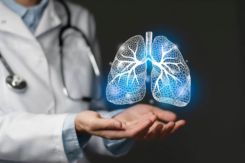
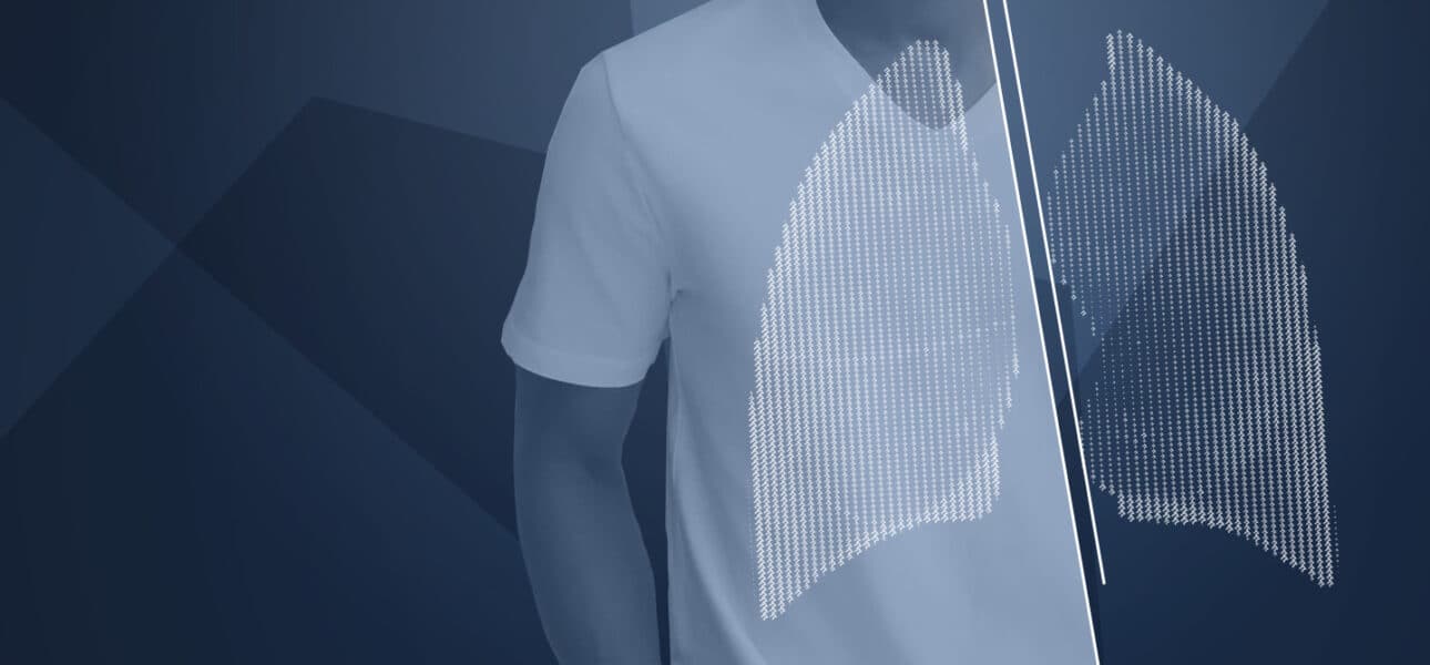
We have all seen a diagram of a lung in our biology textbooks. With the different diagrams, we have been able to learn how it works. However, these inanimate diagrams cannot show how an individual’s organ works, they are only generic models. Today, by digitising these models, it is possible to integrate the data reflecting the organ’s activity. The variables between individuals are thus taken into account thanks to a personalised model. This model, which by definition is more accurate, is called a digital twin.
A digital twin is the digitisation of an object and its environment, which is intended to be true to their physical characteristics. It thus makes it possible to simulate the real behaviour of an object in a virtual environment. It is a model that offers such a high degree of customisation of the digitised version of the object that it is almost as if we were studying the actual object.
The idea is to move towards ever more detailed models, although we know that no model is ever perfect.
This type of numerical simulation is used in many fields of engineering. And it is not difficult to see how such a specific model of an object can be useful in the medical field. After all, when an object can be reduced to a set of equations corresponding to its physical characteristics, everything can be digitised, even the organ of a living being. Martin Genet, a researcher in biomechanics, and several colleagues in the MΞDISIM research team at the Solid Mechanics Laboratory (LMS*) of École Polytechnique (IP Paris)1, are working on personalised lung modelling to study idiopathic pulmonary fibrosis. They believe that the mechanical process of breathing could promote the onset and development of the disease – a kind of mechanical vicious circle. This research is being done with a view to providing doctors with some kind of tool to help them diagnose and manage the patient.
Customising a generic model
First, you must start with the generic model of the target organ. In the case of Martin Genet, this is the lung. Then, using this as a basis, some patient-specific information is applied to customise the generic model. The aim is to create a digital model that corresponds “as closely as possible” to the organ to be studied. “The idea is to move towards ever more detailed models, although we know that no model is ever perfect,” he explains. “A digital twin is therefore a personalised model incorporating the specific data of a patient.”

Even if obtaining a perfect simulation seems impossible, Martin Genet reminds us that perfection is not necessary to obtain relevant medical information. “The perfect model does not exist. A digital twin remains a model, an approximation of reality. So, you can’t reproduce every possible experiment on something real,” he admits. “But a given disease is linked to several physical, chemical, and mechanical processes. Hence, to better understand the disease, and ultimately to better treat it, we obviously must take these processes into account, but it is not necessary to understand all the physico-chemical phenomena that have taken place in the patient’s life,” he insists.
As far as Martin Genet and the MΞDISIM team’s research is concerned, the disease in question is pulmonary fibrosis – a disease that is strongly related to tissue mechanics, corresponding in particular to an overproduction of collagen in lung tissue. As collagen is rigid, too much of it causes the tissue to become rigid. A person with excess collagen will end up forcing his or her breathing, which would in turn aggravate the disease. “This is the fundamental aspect of our research,” explains Martin Genet. “To establish whether mechanical constraints tend to favour the onset and/or the evolution of the disease, and therefore whether the hypothesis of this vicious circle is valid.”
Understanding and classifying fibrosis
Personalised modelling can also be used to go further in diagnosis, particularly for the potential classification of the type of fibrosis the patient has, or simply to monitor and optimise the treatments to be administered. For this, once again, it is not necessary to know the patient’s entire medical history. It could be enough to focus on one aspect of the disease, such as the stiffening of the tissue. Especially since “digital twins of living tissue are now being developed that describe even the interaction of a drug molecule with a subnetwork of a DNA molecule. And all this in a non-invasive way.”
Doctors are very interested in this type of tool because there are very few effective tools for this type of disease today.
The next step is medical diagnosis – although at the research stage it is more appropriate to talk about disease classification. “Among all the types of pulmonary fibrosis, will we be able to classify, quantitatively and objectively, which one the patient in question has?” the researcher asks. If the MΞDISIM team succeeds, the doctor will only have to see the behaviour of the digital lung to determine the appropriate treatment.
The medicine of tomorrow
Moreover, such a tool also has a multitude of other benefits in the health sector, and professionals in this field are aware of this. “Doctors are very interested in this type of biomedical engineering tool,” adds Martin Genet. These tools are intended to facilitate the work of the doctor. Today, there are very few effective tools for this type of disease. Especially since, if this “fundamental aspect” of the research were to validate this “vicious cycle hypothesis”, a digital twin would give the doctor the possibility of predicting the potential development of fibrosis in a patient, and therefore, perhaps, of trying to prevent it.
Finally, one of the major advantages of the digital twin in the medical field lies in the possibility of optimising the treatments administered. Our own organ can be modelled in a simulation that reproduces its functioning and its interaction with other elements, which are themselves modelled. If one of these elements is the drug to be administered, the chemical influence on the organ in question can be simulated. In this way, this digital twin of our organ will give the doctor a quantitative indication of the effectiveness of the treatment to be prescribed (anti-fibrosant, anti-inflammatory, etc.) while allowing them to monitor this treatment in quantitative terms.
“We hope to be able to use the model to describe the evolution of the disease,” summarises the researcher. “Then we can optimise and automate this tool as much as possible, so that it can be easily used in the clinic. The dream would be for the tool to be integrated directly into the hospital’s software pipelines.”


